Introduction
introductionImagine you’re finishing a late evening at the office in Gangnam, staring at your computer for hours, when suddenly you notice something unusual — a streak of flashing light at the edge of your vision, or a new cluster of floaters drifting across your sight like tiny insects. At first, you might think it’s eye strain or fatigue. But for an ophthalmologist, these are red flags: possible signs of a torn retina or even the beginnings of a retinal detachment.
Many patients are surprised to learn that a torn retina and a detached retina are not the same thing. While they are closely related, the difference between the two conditions is critical. A tear may be stabilized with a simple outpatient procedure, while a detachment often requires urgent surgery to save sight. At GS Eye Center in Seoul, we’ve seen how quickly a small retinal tear can evolve into a detachment if left untreated.
This article will walk you through what each condition means, how they develop, and what steps you should take if you ever notice warning signs.
The Retina: Your Eye’s “Projection Screen”
the-retina:-your-eye's-"projection-screen"To understand the difference between a tear and a detachment, it helps to first understand the role of the retina.
The retina is a paper-thin layer of nerve tissue that lines the inside of your eye. Think of it as the projection screen in a theater — it captures incoming light and projects it into electrical signals, which are then transmitted to the brain through the optic nerve. Without a healthy retina, no image can be formed, no matter how clear the cornea or lens in the front of the eye may be.
Because the retina is so delicate, even a small injury can have serious consequences. Tears and detachments are two of the most urgent retinal conditions we see at GS Eye Center. While they sound similar, they represent different stages of damage — with very different treatment implications.
What Is a Retinal Tear?
what-is-a-retinal-tearA retinal tear occurs when a small portion of the thin retinal tissue develops a rip, hole, or break.
How Retinal Tears Develop
how-retinal-tears-developMost tears are caused by changes in the vitreous, the clear gel-like substance that fills the inside of the eye. As we age, the vitreous naturally begins to shrink and pull away from the retina, a process called posterior vitreous detachment (PVD). In many cases, this process is harmless. But sometimes, the vitreous tugs too strongly on the retina and causes a tear.
High myopia, eye trauma, or prior surgery (such as cataract surgery) can make the retina thinner and more vulnerable to these tears.
Symptoms Patients Notice
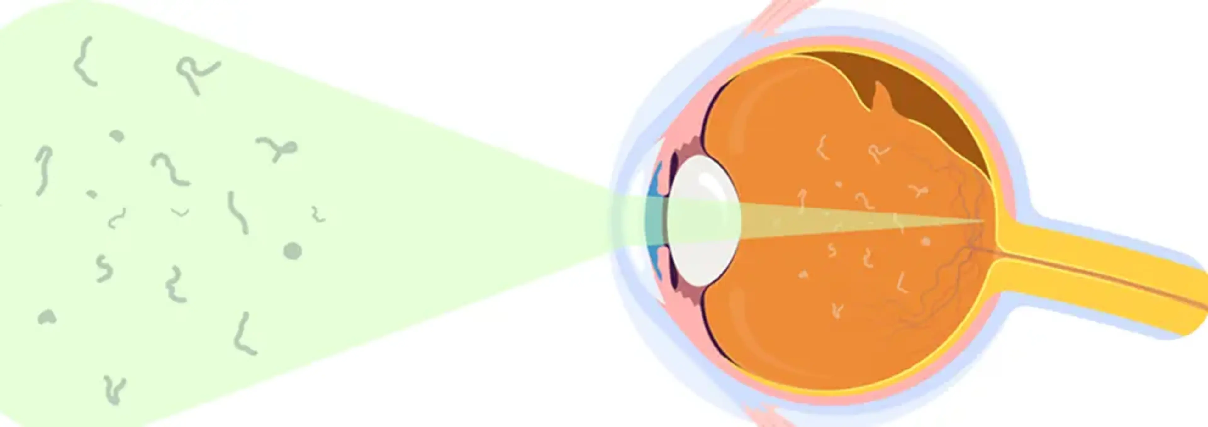
symptoms-patients-noticeFlashes of light (like lightning streaks, especially in dim light)
Sudden new floaters (small dots, cobwebs, or threads drifting across vision)
A shadow or smudge at the edge of vision
It’s important to note that retinal tears may not cause pain, and some patients dismiss the symptoms as fatigue or “just aging.” That’s why awareness is so important.
Why Retinal Tears Are Dangerous
why-retinal-tears-are-dangerousOn their own, retinal tears may not cause significant vision loss. However, they create an opening through which fluid can seep under the retina. This process can separate the retina from the back of the eye — progressing into a retinal detachment.
At GS Eye Center, we often describe a tear as a warning sign. It’s a problem that can usually be treated in under 20 minutes with minimally invasive laser therapy or cryotherapy, but if ignored, it may evolve into something sight-threatening.
What Is a Retinal Detachment?
what-is-a-retinal-detachment
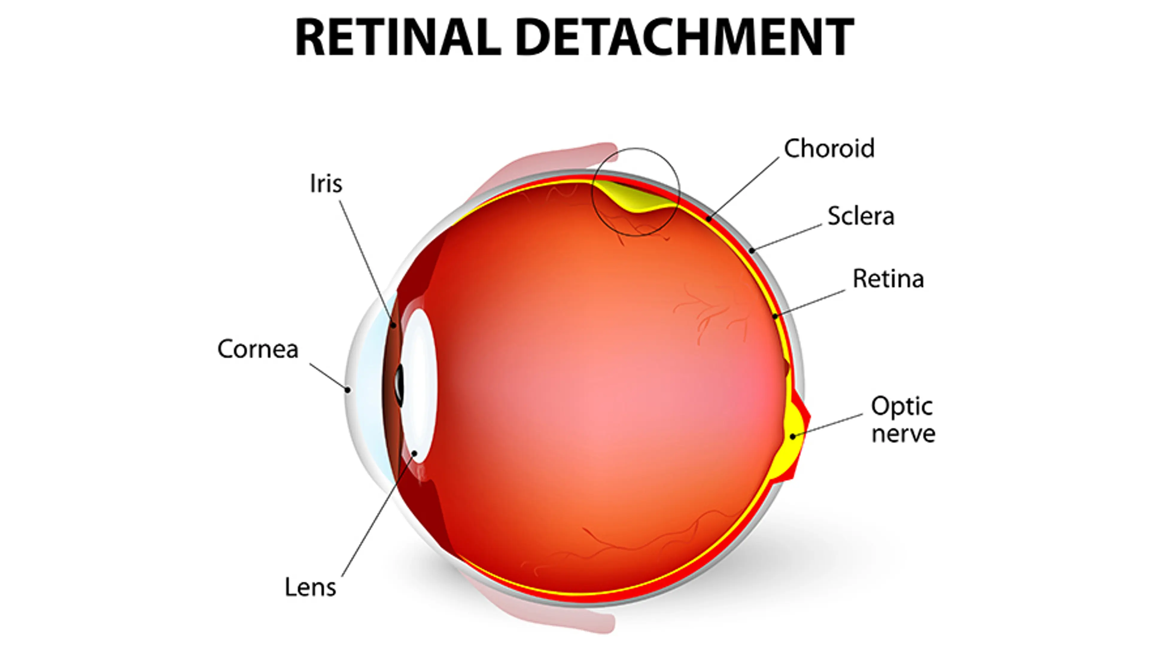
A retinal detachment occurs when part of the retina lifts or peels away from the wall of the eye, like wallpaper separating from plaster.
How Retinal Detachment Progresses
how-retinal-detachment-progressesOnce a tear forms, fluid from inside the eye can seep beneath the retina. This fluid accumulation pushes the retina outward, detaching it from the nourishing tissue layer underneath (the retinal pigment epithelium). Because detached retina cells no longer receive oxygen and nutrients, they quickly begin to lose function.
There are several types of retinal detachment:
Rhegmatogenous detachment (most common): caused by a retinal tear.
Tractional detachment: caused by scar tissue pulling on the retina, often seen in diabetic eye disease.
Exudative detachment: caused by fluid buildup from inflammation, tumors, or vascular disease without a tear.
Symptoms Patients Experience
symptoms-patients-experienceA dark shadow or curtain spreading across the visual field
Blurred or distorted vision in one eye
Loss of side (peripheral) vision
Persistent flashes and floaters (often more intense than in a tear)
Unlike a tear, a detachment typically leads to noticeable vision loss. Left untreated, it can cause permanent blindness in the affected eye.
Treatment Is More Complex
treatment-is-more-complexRetinal detachments require surgery — often within days or even hours of diagnosis. Common procedures include:
Vitrectomy: removal of vitreous gel, with laser sealing and a gas or oil bubble to reattach the retina.
Scleral buckle: a silicone band placed around the eye to relieve traction and hold the retina in place.
Pneumatic retinopexy: a gas bubble injected into the eye to push the retina back against the wall, combined with laser or cryotherapy.
The choice of surgery depends on the location, size, and type of detachment, as well as the patient’s age and eye condition.
Torn Retina vs. Detached Retina: A Simple Analogy
torn-retina-vs.-detached-retina:-a-simple-analogyAn analogy we often share with patients is to imagine the retina as wallpaper inside a room:
A tear is like a small rip in the wallpaper. It may look minor, but it can quickly spread if ignored. Luckily, it can often be patched with a quick repair.
A detachment is when part of the wallpaper has peeled off the wall entirely. At this stage, patching is no longer enough — the wallpaper must be repositioned and secured, a much more involved process.
This analogy helps patients understand why doctors treat retinal tears so urgently, even when vision still seems fine.
Who Is at Risk?
who-is-at-risk
Not everyone faces the same risk of retinal tears or detachments. Factors that increase risk include:
High myopia (severe nearsightedness): In Korea, where myopia is very common, this is one of the strongest risk factors. The elongated shape of the eye stretches the retina, making it thinner and more fragile.
Aging: Most retinal tears occur after age 50, when the vitreous begins to liquefy and pull away.
Previous eye surgery: Patients who have undergone cataract removal, LASIK, or lens implant surgery may face slightly increased risk.
Family history: Genetics can play a role in retinal weakness.
History of retinal detachment in one eye: The other eye is automatically at higher risk.
Eye trauma: Sports injuries, accidents, or blunt trauma can trigger tears.
Systemic diseases: Diabetes and hypertension may increase risk indirectly through effects on retinal blood vessels.
At GS Eye Center, we routinely emphasize preventive screenings for patients who fit these categories, especially those with high myopia — a condition deeply prevalent in urban Korean populations due to intensive near work and lifestyle habits.
Diagnosis: Why Speed Matters
diagnosis:-why-speed-mattersTime is the most critical factor in retinal care. The longer a tear or detachment goes untreated, the more likely permanent vision loss will occur.
Advanced Diagnostics at GS Eye Center
advanced-diagnostics-at-gs-eye-centerWe rely on several technologies to detect and evaluate retinal tears and detachments:
Wide-field retinal imaging: captures up to 200 degrees of the retina in one shot, ideal for spotting peripheral tears.
Optical Coherence Tomography (OCT): provides cross-sectional images of retinal layers, revealing subtle detachments.
Slit-lamp biomicroscopy with special lenses: allows direct, detailed examination of the retina.
What patients often overlook is how much diagnostic testing shapes surgical safety. A careful examination not only confirms the presence of a tear but also reveals whether early detachment has begun — guiding whether immediate outpatient laser treatment or surgery is required.
Treatment Pathways
treatment-pathwaysTreating a Retinal Tear
treating-a-retinal-tearLaser photocoagulation: A laser is used to create small burns around the tear, sealing the retina to underlying tissue.
Cryotherapy: A freezing probe applied to the outer eye wall freezes the tissue around the tear, sealing it shut.
Both are outpatient treatments that typically take less than 20 minutes. Most patients return to normal activities within a day, with minimal discomfort.
Treating a Retinal Detachment
treating-a-retinal-detachmentVitrectomy, scleral buckle, or pneumatic retinopexy are chosen depending on the case.
Recovery is longer, and patients may need to maintain specific head positions to allow a gas bubble to hold the retina in place.
Visual recovery depends heavily on how quickly surgery is performed. In many cases, central vision (macula) can be preserved if the detachment is treated before it spreads there.
Living with Retinal Health in Mind
living-with-retinal-health-in-mindProtecting your retina is not about living in fear, but about being proactive. Some practical steps include:
Don’t ignore symptoms. Flashes, floaters, or sudden shadows should always be checked immediately.
Schedule regular eye exams. Especially if you are highly myopic, over 50, or have had eye surgery.
Choose a clinic with comprehensive retinal services. Look for centers that combine advanced diagnostics with experienced retinal surgeons, so you won’t need to be referred elsewhere if urgent treatment is needed.
At GS Eye Center, our philosophy is patient-first. We often see patients who come in anxious after noticing new floaters, only to discover that the retina is intact. That reassurance is just as valuable as catching a tear early.
Final Thoughts
final-thoughtsA torn retina and a detached retina are not the same — but they are linked. A tear is the warning stage, often manageable with a quick outpatient procedure. A detachment is the emergency stage, requiring urgent surgery to prevent permanent vision loss.
The most important thing for patients to know is this: do not wait. The difference between keeping clear vision and losing it can come down to how quickly you act.
If you’ve noticed sudden floaters, flashes, or shadows in your vision, seek care immediately. At GS Eye Center in Gangnam, our retinal specialists bring more than 20 years of experience, advanced imaging, and surgical precision to protect what matters most — your sight.