Introduction
retina-care:-types-benefits-and-patient-expectationsChoosing the right facility for specialized eye care is a decision that impacts your lifelong independence and quality of life. The retina is a delicate, paper-thin layer of tissue that lines the back of the eye acting much like the film in a camera. When it is damaged the pictures your brain receives become blurry, distorted or disappear entirely. This guide provides a comprehensive roadmap to modern
Retina Care covering advanced diagnostic types the benefits of early intervention and what you should realistically expect as a patient.
Understanding Retina Care: More Than Just a Vision Check
i.-understanding-retina-care:-more-than-just-a-vision-checkTo understand why specialized care is vital, we must first look at how we see.
The Anatomy of Sight
the-anatomy-of-sightThe retina is responsible for converting light into neural signals. It contains millions of light-sensitive cells called photoreceptors (rods and cones). These cells capture light and send signals through the optic nerve to the brain. Without a healthy retina, the brain cannot process the images you see.
When "General" Isn't Enough
when-"general"-isn't-enoughMany patients start their journey at an optometrist for glasses or a general ophthalmologist for cataracts. However, retina care requires a Vitreoretinal Specialist. These are medical doctors who have completed years of additional fellowship training specifically in the diseases and surgery of the vitreous (the gel inside the eye) and the retina.
The "Silent" Window
the-"silent"-windowThe retina is the only place in the body where a doctor can see your blood vessels and nerves directly without surgery. Because of this, retinal health is often the first indicator of systemic issues. Conditions like diabetes, high blood pressure, and even certain autoimmune diseases often show symptoms in the retina long before they affect the rest of the body.
Common Retinal Conditions & Treatment Types
ii.-common-retinal-conditions-and-treatment-types
Retinal diseases vary from age-related wear to sudden emergencies. Below are the most common conditions treated in modern clinics:
Condition | Description | Typical Treatment |
|---|
Macular Degeneration (AMD) | Deterioration of the central part of the retina (the macula). | Anti-VEGF Injections or supplements. |
Diabetic Retinopathy | Damage to blood vessels caused by high blood sugar. | Laser therapy or injections. |
Retinal Detachment | The retina pulls away from the back of the eye. | Emergency Surgery (Vitrectomy or Scleral Buckle). |
Macular Hole | A small break in the macula causing central vision loss. | Vitrectomy with a gas bubble. |
Retinal Vein Occlusion | A stroke in the eye caused by a blocked vein. | Steroid or Anti-VEGF injections. |
Advanced Treatment Angles
advanced-treatment-anglesAnti-VEGF Injections: These medications (like Eylea or Lucentis) stop abnormal blood vessels from leaking fluid, which is the primary cause of vision loss in Wet AMD.
Vitrectomy: A surgical procedure where the vitreous gel is removed to repair a detachment or hole.
Laser Photocoagulation: A precise laser used to seal leaking vessels or tack down retinal tears to prevent a full detachment.
The Benefits of Proactive Retina Care
iii.-the-benefits-of-proactive-retina-careThe single most important factor in retinal health is time.
Vision Preservation: Many retinal diseases such as glaucoma or dry AMD, progress slowly. Regular retina care allows specialists to catch microscopic changes before you notice a blind spot.
Increased Surgical Success: For conditions like retinal detachment, the success rate is significantly higher if the repair happens before the macula (the center of vision) detaches.
Independence: Proactive care ensures you can continue to drive, read and recognize the faces of your loved ones well into your senior years.
Systemic Monitoring: By tracking your retinal blood vessels, your specialist can provide feedback to your primary care doctor about how well your diabetes or blood pressure is being managed.
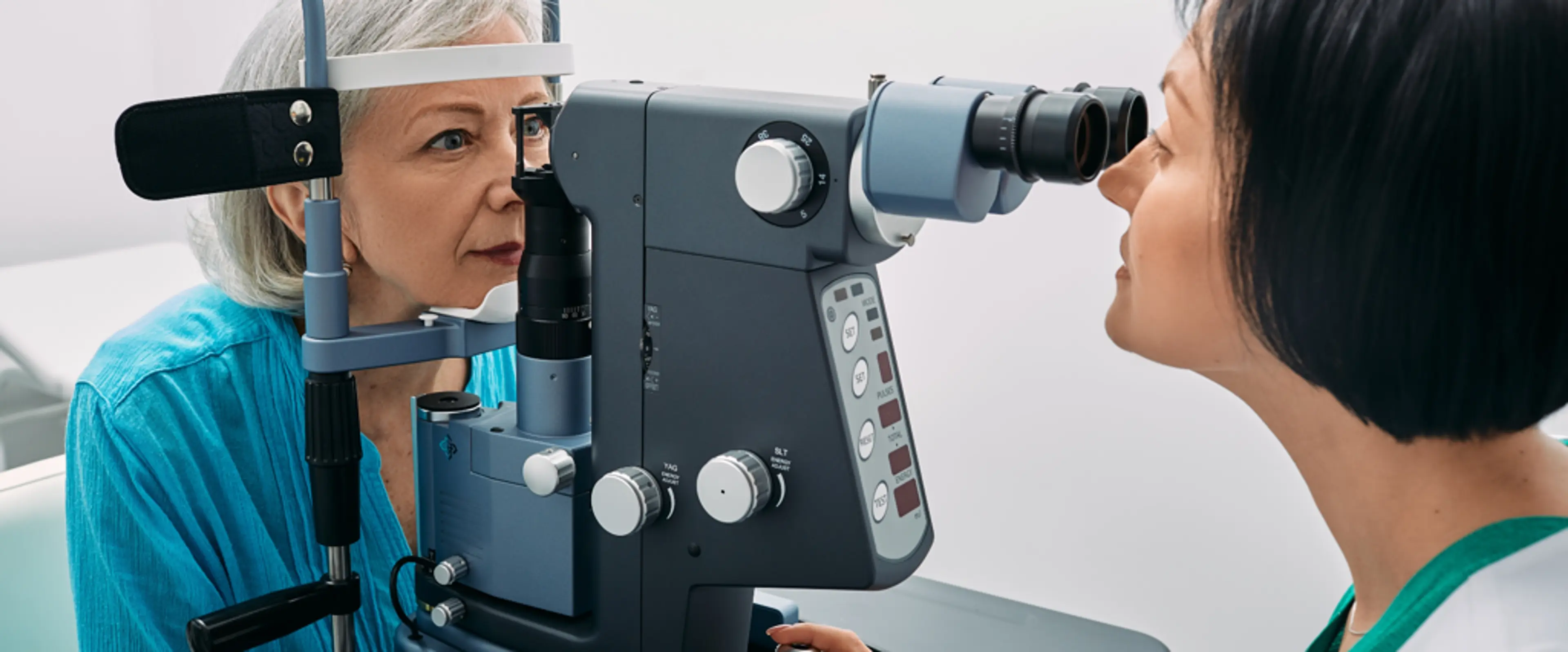
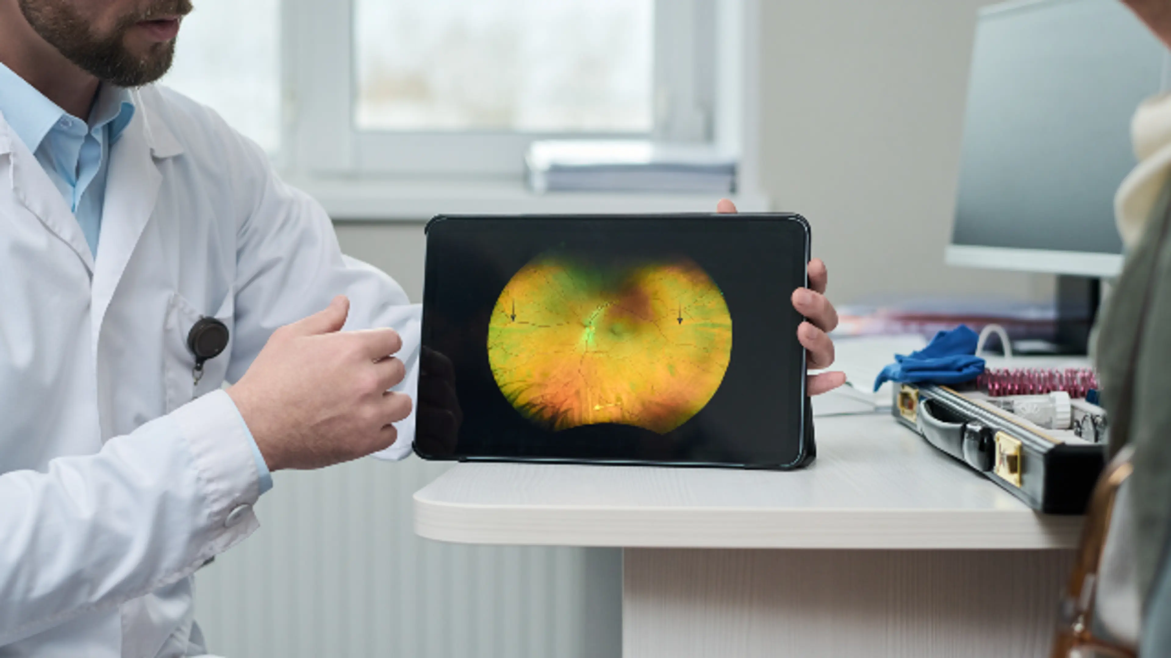
Modern Diagnostic Technologies
iv.-modern-diagnostic-technologiesThe patient experience has been transformed by non-invasive technology. You no longer need to worry about painful tests; most diagnostics are quick and light-based.
Optical Coherence Tomography (OCT): Think of this as a CT scan for the eye. It provides a 3D cross-section of the retina, allowing doctors to see fluid under the surface that is invisible to the naked eye.
Fluorescein Angiography: A specialized dye is injected into the arm to highlight blood flow in the eye. This is essential for mapping diabetic damage.
Fundus Photography: High-resolution digital photos that act as a permanent record to track changes in your eye over several years.
The Global Leader in Eye Care: Why South Korea?
v.-the-global-leader-in-eye-care:-why-south-korea
When searching for the best retina care many patients now look toward South Korea. Known as a global medical hub Seoul offers some of the most advanced ophthalmology clinics in the world. South Korea is widely considered the cheapest and best destination for high-end medical treatment.
Why Choose South Korea?
why-choose-south-koreaTechnology: Korean clinics often house the latest 3D CT and robotic surgical systems before they are widely available in the West.
Expertise: Because of the high volume of patients Korean vitreoretinal surgeons are among the most experienced globally.
Efficiency: Diagnostic tests that take weeks in other countries are often completed in a single afternoon in Seoul.
Cost Comparison: Retina Surgery & Treatments
cost-comparison:-retina-surgery-and-treatmentsProcedure Type | Estimated Cost in South Korea (USD) | Estimated Cost in USA/Europe (USD) |
|---|
Vitrectomy (Standard) | $2,600 – $4,500 | $8,000 – $15,000 |
Retinal Detachment Repair | $2,400 – $5,000 | $10,000 – $18,000 |
Intravitreal Injections (per dose) | $800 – $1,200 | $2,000 – $3,500 |
Laser Photocoagulation | $500 – $1,200 | $2,500 – $5,000 |
Patient Expectations: The Journey to Recovery
vi.-patient-expectations:-the-journey-to-recoveryThe Consultation
the-consultationExpect your eyes to be dilated. This makes your vision blurry and sensitive to light for a few hours, so bring sunglasses and a driver. Your specialist will perform an OCT scan and a thorough physical exam of the eye's interior.
The Recovery Timeline
the-recovery-timelineRecovery from retina surgery is a marathon not a sprint.
Day 1–3: You may experience mild soreness and light sensitivity. If a gas bubble was used your vision in that eye will be like looking through a fishbowl.
Week 1–2: You will use antibiotic and anti-inflammatory drops. You can usually resume light activities like watching TV.
Month 1–6: Vision gradually stabilizes. If a gas bubble was used it will slowly dissolve and be replaced by your eye's natural fluid.
Content Gaps: What Most Guides Miss
vii.-content-gaps:-what-most-guides-missThe Mental Health Aspect
the-mental-health-aspectVision changes can be frightening. It is normal to feel anxiety or frustration during the recovery process. Many top-tier clinics now offer Visual Rehabilitation to help patients adapt to changes while they heal.
Post-Surgical "Face-Down" Positioning
post-surgical-"face-down"-positioningIf you have surgery for a macular hole, you may be asked to remain in a face-down position for several days. This is because the gas bubble must float upward to press against the hole to seal it.
Nutraceuticals (AREDS2)
nutraceuticals-(areds2)For those with Dry AMD, specialized vitamins known as AREDS2 (containing Lutein, Zeaxanthin, Zinc and Vitamin C/E) have been scientifically proven to slow the progression of the disease.
Frequently Asked Questions
viii.-frequently-asked-questions-(faq)1. Is Retina Surgery Painful?
1.-is-retina-surgery-painfulNo. Most procedures are performed under local anesthesia with sedation. You may feel a sensation of pressure but not sharp pain.
2. How Long Until I See Clearly After Surgery?
2.-how-long-until-i-see-clearly-after-surgeryClarity returns over several weeks. If your surgeon used a gas bubble, you won't see clearly until the bubble is at least 50% dissolved.
3. Can I Fly After Retinal Surgery?
3.-can-i-fly-after-retinal-surgeryNo. If a gas bubble was used you cannot fly or travel to high altitudes. The change in pressure can cause the bubble to expand, leading to a dangerous increase in eye pressure.
4. Will I Need Multiple Treatments?
4.-will-i-need-multiple-treatmentsChronic conditions like diabetic retinopathy or Wet AMD often require ongoing injections (every 4–8 weeks) to keep the retina stable.
Conclusion
conclusion